---
description: What does skin cancer look like? View skin cancer pictures to see what to look for. Finding & treating skin cancer early can save your life.
title: Skin Cancer Pictures | What Does Skin Cancer Look Like?
image: https://www.skincancer.org/wp-content/uploads/bannerskincancerpics1.jpg
---
[Home](https://www.skincancer.org/) › [Skin Cancer Information](https://www.skincancer.org/skin-cancer-information/) › Skin Cancer Pictures | What Does Skin Cancer Look Like?
# Skin Cancer Pictures
## What Does Skin Cancer Look Like?
[Skin cancer](https://www.skincancer.org/skin-cancer-information/) can happen to anyone, at any age, on any part of the body. And because skin cancers appear in many shapes and sizes, they can be challenging to identify. While skin cancer pictures can be helpful in learning what skin cancer can look like, getting to know your own skin and understanding [what to look for](https://thebigsee.org/) can help you detect cancer early when it’s easiest to cure.
- [Basal cell carcinoma (BCC) photos](https://www.skincancer.org/skin-cancer-information/skin-cancer-pictures/#bcc): Early and advanced BCCs, and BCCs on various skin tones.
- [Squamous cell carcinoma (SCC) photos](https://www.skincancer.org/skin-cancer-information/skin-cancer-pictures/#scc): Early and late stage SCCs, and SCCs on various skin tones.
- [Actinic keratosis (AK) photos](https://www.skincancer.org/skin-cancer-information/skin-cancer-pictures/#ak): Images of this common precancer.
- [Melanoma photos](https://www.skincancer.org/skin-cancer-information/skin-cancer-pictures/#melanoma): Early stage and late stage melanoma images, and pictures of melanoma on various skin tones.
- [Merkel cell carcinoma photos ](https://www.skincancer.org/skin-cancer-information/skin-cancer-pictures/#mcc)
To catch skin cancer early, you should [examine your skin](https://www.skincancer.org/early-detection/self-exams/) once a month. If you see something [**NEW, CHANGING** OR **UNUSUAL**](https://thebigsee.org/) – even if it looks nothing like what you see in photos – do not wait! Get it checked by a dermatologist right away. Finding and treating skin cancer early can save your life.
**Skin Cancer Image Gallery**
What does cancer look like on skin? Below is a selection of photos that give you a general idea about what skin cancers can look like.
- **Remember that** [skin cancers can look quite different](https://www.skincancer.org/blog/ask-the-expert-are-photos-of-skin-cancer-a-reliable-indicator/) from one person to another due to [skin tone](https://www.skincancer.org/risk-factors/skin-type/), size and type of skin cancer and location on the body.
- **Skin cancer can be tricky** in other ways, too. For example, [melanoma](https://www.skincancer.org/skin-cancer-information/melanoma/) is a type of skin cancer that is often pigmented tan, brown, black, even blue. But [amelanotic melanoma](https://www.skincancer.org/blog/amelanotic-melanoma-it-doesnt-look-like-other-melanomas/) lacks pigment and appears as a skin-tone or pink lesion.
To sum it up, while photos can be helpful, getting your skin examined by a dermatologist is the most vital step in identifying and treating skin cancer.
*Please note: Since not all skin cancers have the same appearance, these photos serve as a general reference for what skin cancer can look like. If you see anything NEW, CHANGING or UNUSUAL on your skin, go get checked by a dermatologist.*
- [Basal Cell Carcinoma Photos](#bcc)
- [Squamous Cell Carcinoma Photos](#scc)
- [Melanoma Photos](#melanoma)
- [Merkel Cell Carcinoma Photos](#mcc)
- [Actinic Keratosis](#ak)
- [Rare Skin Cancers](#rare)
- [Newly Diagnosed with Skin Cancer](https://www.skincancer.org/skin-cancer-information/newly-diagnosed-with-skin-cancer/)
### Basal Cell Carcinoma (BCC) Photos
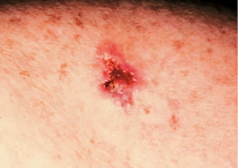
*An open sore that does not heal. (BCC)*

*An open sore that bleeds, oozes, or crusts. (BCC)*

*A reddish patch or irritated area. (BCC)*

*A shiny bump or nodule. (BCC)*

*White, yellow or waxy scar. (BCC)*

*A basal cell carcinoma may be pigmented on skin of color.*Photo: Andrew Alexis, MD, MPH**

*BCC on the nose.*

*A small pink growth with a slightly raised, rolled edge and a crusted indentation in the center. (BCC)*

*A shiny bump or nodule. (BCC)*

*Pigmented BCC behind the ear. *Photo: Hugh Gloster, MD**

*A scar-like area that is white, yellow or waxy, and often has poorly defined borders. (BCC)*

*Pigmented BCC. *Photo credit: Hugh Gloster, MD**

*BCC on the nose. *Photo: Hugh Gloster, MD**

*A pink growth. (BCC)*

*A subtle pearly lesion in the middle of the lower eyelid’s lash line is a BCC.*

*Basal cell carcinoma presenting as a pink growth. *Photo: International Skin Imaging Collaboration at isic-archive.com**

*Basal cell carcinoma presenting as an open sore on the ear. *Photo: International Skin Imaging Collaboration at isic-archive.com**

*Basal cell carcinoma on the leg. *Photo: International Skin Imaging Collaboration at isic-archive.com**

*Basal cell carcinoma on the posterior torso. *Photo: International Skin Imaging Collaboration at isic-archive.com**

*Basal cell carcinoma on the neck. *Photo: International Skin Imaging Collaboration at isic-archive.com**

*Basal cell carcinoma on the anterior torso. *Photo: International Skin Imaging Collaboration at isic-archive.com**

*Basal cell carcinoma on the anterior torso. *Photo: International Skin Imaging Collaboration at isic-archive.com**

*Basal cell carcinoma on the anterior torso. *Photo: International Skin Imaging Collaboration at isic-archive.com**
For information on basal cell carcinoma signs and symptoms, visit our **[BCC Warning Signs](https://www.skincancer.org/skin-cancer-information/basal-cell-carcinoma/bcc-warning-signs-images/#five)** page.
#### Basal Cell Carcinoma (BCC) Case Study
**Inconvenient BCC, Good Outcome**
*Deborah S. Sarnoff, MD*
A patient in his 40s with the genetic condition known as Gorlin syndrome had been treated for many basal cell carcinomas. During the COVID pandemic, he noticed a new bump on his upper lip. It didn’t seem serious to him, so, like many patients during that time, he did not see his dermatologist. When he finally did get checked, it was indeed a BCC and had grown deep enough to require several stages of Mohs surgery. The surgery eliminated the cancer but left the lower part of his face disfigured (top photo). Dr. Sarnoff’s business partner and husband, plastic surgeon Robert H. Gotkin, MD, was able to reconstruct the area with minimal scarring and an excellent cosmetic result (bottom photo, after healing). Read the full story here: https://www.skincancer.org/blog/we-dont-want-to-scare-you-but/

### Squamous Cell Carcinoma (SCC) Images

*A persistent, scaly red patch with irregular borders that sometimes crusts or bleeds. (SCC)*

*An elevated growth with a central depression that occasionally bleeds. It may rapidly increase in size. (SCC)*

*An open sore that bleeds and crusts and persists for weeks. (SCC)*
![crusty spot eye squamous cell carcinoma]()
*This crusty spot near the eye was diagnosed as a squamous cell carcinoma (SCC).*

*A wart-like growth that crusts and occasionally bleeds. (SCC)*
![cutaneous horn squamous cell carcinoma lower eyelid]()
*This horn-shaped growth on the lower eyelid, known as a cutaneous horn, is an SCC.*

*An open sore that bleeds or crusts and persists for weeks. (SCC)*

*A persistent, scaly red patch with irregular borders that sometimes crusts or bleeds. (SCC)*

*Squamous cell carcinoma on the scalp. *Photo: Hugh Gloster, MD**

*Large squamous cell carcinoma*

*Squamous cell carcinoma on the nose. *Photo: International Skin Imaging Collaboration at isic-archive.com**

*Squamous cell carcinoma on the head. *Photo: International Skin Imaging Collaboration at isic-archive.com**

*Squamous cell carcinoma on the neck. *Photo: International Skin Imaging Collaboration at isic-archive.com**

*Squamous cell carcinoma.*Photo: International Skin Imaging Collaboration at isic-archive.com**

*Squamous cell carcinoma on the head. *Photo: International Skin Imaging Collaboration at isic-archive.com**
For information on squamous cell carcinoma signs and symptoms, visit our **[SCC Warning Signs](https://www.skincancer.org/skin-cancer-information/squamous-cell-carcinoma/scc-warning-signs-and-images/)** page.
#### Squamous Cell Carcinoma (SCC) Case Study
**A Large Squamous Cell Carcinoma**
*Deborah S. Sarnoff, MD*
Before being admitted to an assisted living facility, a older woman was required to have a thorough physical. The doctor (and her adult children who had not seen her undressed in years), were shocked to see a large, raised open wound on her left shoulder and back. For years, she had been covering it and hiding it under clothes. She didn’t want to rock the boat or make a fuss. The lesion (top photo) turned out to be a large SCC, the second most common type of skin cancer. The tumor required extensive surgery followed by radiation. The patient was extremely lucky: Her cancer was eliminated, and Dr. Sarnoff’s business partner and husband Robert H. Gotkin, MD, expertly closed the wound (bottom photo). Dr. Sarnoff strongly advocates robust treatment for older people with skin cancer. The earlier it is diagnosed, the less onerous the treatment, and it can make a huge difference in a person’s quality of life.

### Melanoma Photos

*Asymmetrical melanoma.*

*The borders of an early melanoma tend to be uneven. The edges may be scalloped or notched.*

*Having a mole with a variety of colors is a melanoma warning sign.*

*Melanomas usually are larger in diameter but may be smaller when first detected.*
![stage 1a melanoma]()
*Stage IA melanoma.*

*Subungual melanoma under the nail.*

*Melanoma evolved and grew outward, upward and inward in a matter of months.*

*Acral lentiginous melanoma.*

*Nodular melanoma 1.3mm on upper back. *Photo: Elizabeth K. Hale, MD**

*Superficial melanoma 0.55mm on upper arm. *Photo: Elizabeth K. Hale, MD**

*Amelanotic melanomas may be pinkish-looking, reddish, purple, normal skin color or essentially clear and colorless.*

*Acral lentiginous melanoma.*

*Amelanotic melanoma.*

*Dark melanoma, 4 mms. *Photo: International Skin Imaging Collaboration at isic-archive.com**

*Dark melanoma, 4 mms. *Photo: International Skin Imaging Collaboration at isic-archive.com**

*Dark melanoma, 4 mms. *Photo: International Skin Imaging Collaboration at isic-archive.com**

*Melanoma, 1.3mm, right posterior shoulder. *Photo: Sarah Gee, MD**

*Melanoma in situ 1 cm on the right calf. *Photo: Jane Yoo, MD**

*Melanoma *Photo: International Skin Imaging Collaboration**

*Melanoma *Photo: International Skin Imaging Collaboration**

*Melanoma *Photo: International Skin Imaging Collaboration**

*Melanoma *Photo: International Skin Imaging Collaboration**

*Melanoma *Photo: International Skin Imaging Collaboration**

*Melanoma *Photo: International Skin Imaging Collaboration**

*Melanoma *Photo: International Skin Imaging Collaboration**

*Nodular melanoma on the leg of a Native American woman.
*Photo: International Skin Imaging Collaboration**
For information on melanoma signs and symptoms, visit our **[Melanoma Warning Signs](https://www.skincancer.org/skin-cancer-information/melanoma/melanoma-warning-signs-and-images/)** page.
#### Melanoma Case Study
**Not a “Boil” but an Advanced Melanoma **
*Deborah S. Sarnoff, MD*
The power of denial can be strong. This man believed the large, dark growth on his back was a boil, despite oozing and bleeding for months. He kept bandaging it and thought it was just slow to heal. When he was finally referred to Dr. Sarnoff, a biopsy and other tests revealed a melanoma so large and deep that it had already spread to the man’s liver and brain. He was referred to an oncologist and began an immunotherapy regimen that did not appear to be working, which happens with some patients. (Thanks to recent innovations, there may be other treatment options, including participation in a clinical trial.) This is a powerful reminder of the importance of early detection.

### Merkel Cell Carcinoma (MCC) Pictures

*Merkel cell carcinoma on woman's left eyelid. *Photos: Paul Nghiem, MD, PhD**

*Merkel cell carcinoma on the arm. *Photos: Paul Nghiem, MD, PhD**

*Merkel cell carcinoma on forehead and arm. *Photos: Paul Nghiem, MD, PhD**

*Merkel cell carcinoma on the lower leg. *Photos: Paul Nghiem, MD, PhD**

*Bump on leg that turned out to be merkel cell carcinoma. *Photos: Paul Nghiem, MD, PhD**
For information on Merkel cell carcinoma signs and symptoms, visit our **[MCC Warning Signs](https://www.skincancer.org/skin-cancer-information/merkel-cell-carcinoma/mcc-warning-signs/)** page.
### Actinic Keratosis
Actinic keratosis (also known as solar keratosis) is the most common precancer. For more photos and information on actinic keratosis warning signs and symptoms, visit our **[Actinic Keratosis Warning Signs](https://www.skincancer.org/skin-cancer-information/actinic-keratosis/actinic-keratosis-warning-signs-and-images/)** page.
- ![]()
- Actinic keratoses located on the hand.
- ![]()
- Actinic keratoses located on the ear.
- ![]()
- Actinic keratoses located on the hand.
- ![Actinic keratoses located on the scalp.]()
- Actinic keratoses located on the scalp.
- ![Actinic keratoses located on the ear.]()
- Actinic keratoses located on the ear.
- ![Actinic keratoses located on the scalp.]()
- Actinic keratoses located on the scalp.
All Actinic Keratosis Basal Cell Carcinoma Melanoma Merkel Cell Carcinoma Squamous Cell Carcinoma
### Actinic Keratosis (AK) Photos
![Actinic keratosis on head or neck. Photo: International Skin Imaging Collaboration at isic-archive.com]()
Actinic keratosis on head or neck. Photo: International Skin Imaging Collaboration at isic-archive.com
![Actinic keratosis. Photo: International Skin Imaging Collaboration at isic-archive.com]()
Actinic keratosis. Photo: International Skin Imaging Collaboration at isic-archive.com
![ Actinic keratosis on posterior torso. Photo: International Skin Imaging Collaboration at isic-archive.com]()
Actinic keratosis on posterior torso. Photo: International Skin Imaging Collaboration at isic-archive.com
![Actinic keratosis on head or neck. Photo: International Skin Imaging Collaboration at isic-archive.com]()
Actinic keratosis on head or neck. Photo: International Skin Imaging Collaboration at isic-archive.com
![Actinic keratoses on scalp. Photo: Almirall, SA]()
Actinic keratoses on scalp. Photo: Almirall, SA
![Close up of actinic keratoses on scalp. Photo: Almirall, SA]()
Close up of actinic keratoses on scalp. Photo: Almirall, SA
![Actinic keratoses on scalp. Photo: Almirall, SA]()
Actinic keratoses on scalp. Photo: Almirall, SA
![Multiple actinic keratoses on scalp. Photo: Almirall, SA]()
Multiple actinic keratoses on scalp. Photo: Almirall, SA
![Actinic keratoses located on the scalp.]()
Actinic keratoses located on the scalp.
![Actinic keratoses located on the scalp.]()
Actinic keratoses located on the scalp.
![Actinic keratoses located on the ear.]()
Actinic keratoses located on the ear.
![Actinic keratoses located on the hand.]()
Actinic keratoses located on the hand.
![Actinic keratoses located on the ear.]()
Actinic keratoses located on the ear.
![Actinic keratoses located on the hand.]()
Actinic keratoses located on the hand.
For information on Actinic Keratosis signs and symptoms, visit our **[AK Warning Signs](https://www.skincancer.org/skin-cancer-information/actinic-keratosis/actinic-keratosis-warning-signs-and-images/)** page.
### Basal Cell Carcinoma (BCC) Photos
![Morpheaform BCC on a Black woman’s face]()
Morpheaform BCC on a Black woman’s face
![Basal cell carcinoma, shiny bump above upper lip \
Photo credit: Richard P. Usatine, MD.
]()
Basal cell carcinoma, shiny bump above upper lip
Photo credit: Richard P. Usatine, MD.
![Basal cell carcinoma \
Photo credit: Richard P. Usatine, MD.
]()
Basal cell carcinoma
Photo credit: Richard P. Usatine, MD.
![Large basal cell carcinoma \
Photo credit: Richard P. Usatine, MD.]()
Large basal cell carcinoma
Photo credit: Richard P. Usatine, MD.
![Basal cell carcinoma on ear \
Photo credit: Richard P. Usatine, MD.]()
Basal cell carcinoma on ear
Photo credit: Richard P. Usatine, MD.
![Large basal cell carcinoma on scalp \
Photo credit: Richard P. Usatine, MD.]()
Large basal cell carcinoma on scalp
Photo credit: Richard P. Usatine, MD.
![Pigmented basal cell carcinoma on scalp \
Photo credit: Richard P. Usatine, MD.]()
Pigmented basal cell carcinoma on scalp
Photo credit: Richard P. Usatine, MD.
![Pigmented basal cell carcinoma on cheek \
Photo credit: Richard P. Usatine, MD.]()
Pigmented basal cell carcinoma on cheek
Photo credit: Richard P. Usatine, MD.
![Basal cell carcinoma - shiny bump \
Photo credit: Richard P. Usatine, MD.]()
Basal cell carcinoma - shiny bump
Photo credit: Richard P. Usatine, MD.
![Basal cell carcinoma - a sore that does not heal \
Photo credit: Richard P. Usatine, MD.]()
Basal cell carcinoma - a sore that does not heal
Photo credit: Richard P. Usatine, MD.
![Basal cell carcinoma on cheek \
Photo credit: Richard P. Usatine, MD.]()
Basal cell carcinoma on cheek
Photo credit: Richard P. Usatine, MD.
![Large, pigmented basal cell carcinoma \
Photo credit: Richard P. Usatine, MD.]()
Large, pigmented basal cell carcinoma
Photo credit: Richard P. Usatine, MD.
![Basal cell carcinoma - a red, irritated patch \
Photo credit: Richard P. Usatine, MD.]()
Basal cell carcinoma - a red, irritated patch
Photo credit: Richard P. Usatine, MD.
![Basal cell carcinoma on the posterior torso. \Photo: International Skin Imaging Collaboration at isic-archive.com\]()
Basal cell carcinoma on the posterior torso. *Photo: International Skin Imaging Collaboration at isic-archive.com*
![Basal cell carcinoma on the neck. \Photo: International Skin Imaging Collaboration at isic-archive.com\]()
Basal cell carcinoma on the neck. *Photo: International Skin Imaging Collaboration at isic-archive.com*
![Basal cell carcinoma on the anterior torso. \Photo: International Skin Imaging Collaboration at isic-archive.com\]()
Basal cell carcinoma on the anterior torso. *Photo: International Skin Imaging Collaboration at isic-archive.com*
![Basal cell carcinoma on the anterior torso. \Photo: International Skin Imaging Collaboration at isic-archive.com\]()
Basal cell carcinoma on the anterior torso. *Photo: International Skin Imaging Collaboration at isic-archive.com*
![Basal cell carcinoma on the anterior torso. \Photo: International Skin Imaging Collaboration at isic-archive.com\]()
Basal cell carcinoma on the anterior torso. *Photo: International Skin Imaging Collaboration at isic-archive.com*
![Basal cell carcinoma presenting as an open sore on the ear. \Photo: International Skin Imaging Collaboration at isic-archive.com\]()
Basal cell carcinoma presenting as an open sore on the ear. *Photo: International Skin Imaging Collaboration at isic-archive.com*
![Basal cell carcinoma presenting as a pink growth. \Photo: International Skin Imaging Collaboration at isic-archive.com\]()
Basal cell carcinoma presenting as a pink growth. *Photo: International Skin Imaging Collaboration at isic-archive.com*
![Basal cell carcinoma on the leg. \Photo: International Skin Imaging Collaboration at isic-archive.com\]()
Basal cell carcinoma on the leg. *Photo: International Skin Imaging Collaboration at isic-archive.com*
![BCC on the nose.]()
BCC on the nose.
![Pigmented BCC. \Photo credit: Hugh Gloster, MD\]()
Pigmented BCC. *Photo credit: Hugh Gloster, MD*
![BCC on the nose.
\Photo: Hugh Gloster, MD\]()
BCC on the nose. *Photo: Hugh Gloster, MD*
![Pigmented BCC behind the ear. \Photo: Hugh Gloster, MD\]()
Pigmented BCC behind the ear. *Photo: Hugh Gloster, MD*
![A small pink growth with a slightly raised, rolled edge and a crusted indentation in the center. (BCC) ]()
A small pink growth with a slightly raised, rolled edge and a crusted indentation in the center. (BCC)
![A basal cell carcinoma may be pigmented on skin of color.\Photo: Andrew Alexis, MD, MPH\]()
A basal cell carcinoma may be pigmented on skin of color.*Photo: Andrew Alexis, MD, MPH*
![A scar-like area that is white, yellow or waxy, and often has poorly defined borders. (BCC) ]()
A scar-like area that is white, yellow or waxy, and often has poorly defined borders. (BCC)
![A pink growth. (BCC) ]()
A pink growth. (BCC)
![A reddish patch or irritated area. (BCC)]()
A reddish patch or irritated area. (BCC)
![A shiny bump or nodule. (BCC) ]()
A shiny bump or nodule. (BCC)
![An open sore that bleeds, oozes, or crusts. (BCC)]()
An open sore that bleeds, oozes, or crusts. (BCC)
![White, yellow or waxy scar. (BCC)]()
White, yellow or waxy scar. (BCC)
![An open sore that does not heal. (BCC)]()
An open sore that does not heal. (BCC)
![A shiny bump or nodule. (BCC)]()
A shiny bump or nodule. (BCC)
![A subtle pearly lesion in the middle of the lower eyelid’s lash line is a BCC.]()
A subtle pearly lesion in the middle of the lower eyelid’s lash line is a BCC.
![]()
**Basal Cell Carcinoma (BCC) Case Study**
Inconvenient BCC, Good Outcome
Deborah S. Sarnoff, MD
A patient in his 40s with the genetic condition known as Gorlin syndrome had been treated for many basal cell carcinomas. During the COVID pandemic, he noticed a new bump on his upper lip. It didn’t seem serious to him, so, like many patients during that time, he did not see his dermatologist. When he finally did get checked, it was indeed a BCC and had grown deep enough to require several stages of Mohs surgery. The surgery eliminated the cancer but left the lower part of his face disfigured (top photo). Dr. Sarnoff’s business partner and husband, plastic surgeon Robert H. Gotkin, MD, was able to reconstruct the area with minimal scarring and an excellent cosmetic result (bottom photo, after healing). Read the full story here: https://skincancer.org/blog/we-dont-want-to-scare-you-but/
For information on Basal Cell Carcinoma signs and symptoms, visit our **[BCC Warning Signs](https://www.skincancer.org/skin-cancer-information/basal-cell-carcinoma/bcc-warning-signs-images/#five)** page.
### Melanoma Photos
![Amelanotic melanoma on the temple. Photo: Deborah S. Sarnoff, MD]()
Amelanotic melanoma on the temple. Photo: Deborah S. Sarnoff, MD
![Stage II acral lentiginous melanoma on left foot. Photo courtesy of: Dorothy Overstreet]()
Stage II acral lentiginous melanoma on left foot. Photo courtesy of: Dorothy Overstreet
![Superficial melanoma 0.55mm Upper Arm \
Credit: Elizabeth K. Hale, MD]()
Superficial melanoma 0.55mm Upper Arm
Credit: Elizabeth K. Hale, MD
![Nodular melanoma 1.3mm upper back \
Credit: Elizabeth K. Hale, MD]()
Nodular melanoma 1.3mm upper back
Credit: Elizabeth K. Hale, MD
![Subungual melanoma]()
Subungual melanoma
![Melanoma on the lip of a Black man]()
Melanoma on the lip of a Black man
![Melanoma on the eyebrow of a Hispanic man]()
Melanoma on the eyebrow of a Hispanic man
![ Large melanoma with varied colors \
Photo credit: Richard P. Usatine, MD.]()
Large melanoma with varied colors
Photo credit: Richard P. Usatine, MD.
![ Dark, raised melanoma with irregular borders\
Photo credit: Richard P. Usatine, MD.]()
Dark, raised melanoma with irregular borders
Photo credit: Richard P. Usatine, MD.
![Large melanoma with irregular borders\
Photo credit: Richard P. Usatine, MD.]()
Large melanoma with irregular borders
Photo credit: Richard P. Usatine, MD.
![Melanoma with asymmetry and irregular borders \
Photo credit: Richard P. Usatine, MD.
]()
Melanoma with asymmetry and irregular borders
Photo credit: Richard P. Usatine, MD.
![Acral lentiginous melanoma on heel \
Photo credit: Richard P. Usatine, MD.
]()
Acral lentiginous melanoma on heel
Photo credit: Richard P. Usatine, MD.
![Melanoma on cheek\
Photo credit: Richard P. Usatine, MD.
]()
Melanoma on cheek
Photo credit: Richard P. Usatine, MD.
![Dark, raised melanoma\
Photo credit: Richard P. Usatine, MD.]()
Dark, raised melanoma
Photo credit: Richard P. Usatine, MD.
![Melanoma, irregular borders\
Photo credit: Richard P. Usatine, MD.]()
Melanoma, irregular borders
Photo credit: Richard P. Usatine, MD.
![Subungual melanoma under toenail\
Photo credit: Richard P. Usatine, MD.]()
Subungual melanoma under toenail
Photo credit: Richard P. Usatine, MD.
![Subungual melanoma on fingernail \
Photo credit: Richard P. Usatine, MD.]()
Subungual melanoma on fingernail
Photo credit: Richard P. Usatine, MD.
![Melanoma on finger\
Photo credit: Richard P. Usatine, MD.]()
Melanoma on finger
Photo credit: Richard P. Usatine, MD.
![Acral lentiginous melanoma on heel of foot \
Photo credit: Richard P. Usatine, MD.
]()
Acral lentiginous melanoma on heel of foot
Photo credit: Richard P. Usatine, MD.
![Melanoma evolved and grew outward, upward and inward in a matter of months.]()
Melanoma evolved and grew outward, upward and inward in a matter of months.
![Melanoma \Photo: International Skin Imaging Collaboration\]()
Melanoma *Photo: International Skin Imaging Collaboration*
![Melanoma \Photo: International Skin Imaging Collaboration\]()
Melanoma *Photo: International Skin Imaging Collaboration*
![Melanoma \Photo: International Skin Imaging Collaboration\]()
Melanoma *Photo: International Skin Imaging Collaboration*
![Melanoma \Photo: International Skin Imaging Collaboration\]()
Melanoma *Photo: International Skin Imaging Collaboration*
![Melanoma \Photo: International Skin Imaging Collaboration\]()
Melanoma *Photo: International Skin Imaging Collaboration*
![Melanoma \Photo: International Skin Imaging Collaboration\]()
Melanoma *Photo: International Skin Imaging Collaboration*
![Melanoma \Photo: International Skin Imaging Collaboration\]()
Melanoma *Photo: International Skin Imaging Collaboration*
![Nodular melanoma on the leg of a Native American woman.
\
\Photo: International Skin Imaging Collaboration\]()
Nodular melanoma on the leg of a Native American woman.
*Photo: International Skin Imaging Collaboration*
![Melanoma in situ 1 cm on the right calf.
\Photo: Jane Yoo, MD\]()
Melanoma in situ 1 cm on the right calf. *Photo: Jane Yoo, MD*
![Melanoma, 1.3mm, right posterior shoulder.
\Photo: Sarah Gee, MD\]()
Melanoma, 1.3mm, right posterior shoulder. *Photo: Sarah Gee, MD*
![Dark melanoma, 4 mms.
\Photo: International Skin Imaging Collaboration at isic-archive.com\]()
Dark melanoma, 4 mms. *Photo: International Skin Imaging Collaboration at isic-archive.com*
![Dark melanoma, 4 mms.
\Photo: International Skin Imaging Collaboration at isic-archive.com\]()
Dark melanoma, 4 mms. *Photo: International Skin Imaging Collaboration at isic-archive.com*
![Dark melanoma, 4 mms.
\Photo: International Skin Imaging Collaboration at isic-archive.com\]()
Dark melanoma, 4 mms. *Photo: International Skin Imaging Collaboration at isic-archive.com*
![Superficial melanoma 0.55mm on upper arm. \Photo: Elizabeth K. Hale, MD\]()
Superficial melanoma 0.55mm on upper arm. *Photo: Elizabeth K. Hale, MD*
![Nodular melanoma 1.3mm on upper back. \Photo: Elizabeth K. Hale, MD\]()
Nodular melanoma 1.3mm on upper back. *Photo: Elizabeth K. Hale, MD*
![Acral lentiginous melanoma.]()
Acral lentiginous melanoma.
![Acral lentiginous melanoma.]()
Acral lentiginous melanoma.
![Amelanotic melanomas may be pinkish-looking, reddish, purple, normal skin color or essentially clear and colorless.]()
Amelanotic melanomas may be pinkish-looking, reddish, purple, normal skin color or essentially clear and colorless.
![Amelanotic melanoma.]()
Amelanotic melanoma.
![Asymmetrical melanoma.
]()
Asymmetrical melanoma.
![The borders of an early melanoma tend to be uneven. The edges may be scalloped or notched.]()
The borders of an early melanoma tend to be uneven. The edges may be scalloped or notched.
![Melanomas usually are larger in diameter but may be smaller when first detected.]()
Melanomas usually are larger in diameter but may be smaller when first detected.
![Having a mole with a variety of colors is a melanoma warning sign.]()
Having a mole with a variety of colors is a melanoma warning sign.
![Subungual melanoma under the nail.]()
Subungual melanoma under the nail.
![Stage IA melanoma.]()
Stage IA melanoma.
![]()
**Melanoma Case Study**
Not a “Boil” but an Advanced Melanoma
Deborah S. Sarnoff, MD
The power of denial can be strong. This man believed the large, dark growth on his back was a boil, despite oozing and bleeding for months. He kept bandaging it and thought it was just slow to heal. When he was finally referred to Dr. Sarnoff, a biopsy and other tests revealed a melanoma so large and deep that it had already spread to the man’s liver and brain. He was referred to an oncologist and began an immunotherapy regimen that did not appear to be working, which happens with some patients. (Thanks to recent innovations, there may be other treatment options, including participation in a clinical trial.) This is a powerful reminder of the importance of early detection.
For information on Melanoma signs and symptoms, visit our **[Melanoma Warning Signs](https://www.skincancer.org/skin-cancer-information/melanoma/melanoma-warning-signs-and-images/)** page.
### Merkel Cell Carcinoma (MCC) Pictures
![Merkel cell carcinoma on the arm. \Photos: Paul Nghiem, MD, PhD\]()
Merkel cell carcinoma on the arm. *Photos: Paul Nghiem, MD, PhD*
![Merkel cell carcinoma on woman's left eyelid. \Photos: Paul Nghiem, MD, PhD\]()
Merkel cell carcinoma on woman's left eyelid. *Photos: Paul Nghiem, MD, PhD*
![Merkel cell carcinoma on the lower leg. \Photos: Paul Nghiem, MD, PhD\]()
Merkel cell carcinoma on the lower leg. *Photos: Paul Nghiem, MD, PhD*
![Bump on leg that turned out to be merkel cell carcinoma. \Photos: Paul Nghiem, MD, PhD\]()
Bump on leg that turned out to be merkel cell carcinoma. *Photos: Paul Nghiem, MD, PhD*
![Merkel cell carcinoma on forehead and arm. \Photos: Paul Nghiem, MD, PhD\]()
Merkel cell carcinoma on forehead and arm. *Photos: Paul Nghiem, MD, PhD*
For information on Merkel Cell Carcinoma signs and symptoms, visit our **[MCC Warning Signs](https://www.skincancer.org/skin-cancer-information/merkel-cell-carcinoma/mcc-warning-signs/)** page.
### Squamous Cell Carcinoma (SCC) Photos
![Bowen's disease (SCC in situ) on the finger of a Black man]()
Bowen's disease (SCC in situ) on the finger of a Black man
![Squamous cell carcinoma, scaly patch on cheek\
Photo credit: Richard P. Usatine, MD.]()
Squamous cell carcinoma, scaly patch on cheek
Photo credit: Richard P. Usatine, MD.
![Squamous cell carcinoma on light skin \
Photo credit: Richard P. Usatine, MD.]()
Squamous cell carcinoma on light skin
Photo credit: Richard P. Usatine, MD.
![Squamous cell carcinoma on ear \
Photo credit: Richard P. Usatine, MD.]()
Squamous cell carcinoma on ear
Photo credit: Richard P. Usatine, MD.
![Squamous cell carcinoma on forearm of man with darker skin tone \
Photo credit: Richard P. Usatine, MD.]()
Squamous cell carcinoma on forearm of man with darker skin tone
Photo credit: Richard P. Usatine, MD.
![Raised, reddish squamous cell carcinoma \
Photo credit: Richard P. Usatine, MD.]()
Raised, reddish squamous cell carcinoma
Photo credit: Richard P. Usatine, MD.
![Squamous cell carcinoma near hairline \
Photo credit: Richard P. Usatine, MD.]()
Squamous cell carcinoma near hairline
Photo credit: Richard P. Usatine, MD.
![Large, crusty squamous cell carcinoma on ear \
Photo credit: Richard P. Usatine, MD.]()
Large, crusty squamous cell carcinoma on ear
Photo credit: Richard P. Usatine, MD.
![Large squamous cell carcinoma on lip \
Photo credit: Richard P. Usatine, MD.]()
Large squamous cell carcinoma on lip
Photo credit: Richard P. Usatine, MD.
![Squamous cell carcinoma beside toenail \
Photo credit: Richard P. Usatine, MD.]()
Squamous cell carcinoma beside toenail
Photo credit: Richard P. Usatine, MD.
![Large, crusty squamous cell carcinoma \
Photo credit: Richard P. Usatine, MD.]()
Large, crusty squamous cell carcinoma
Photo credit: Richard P. Usatine, MD.
![Raised, red, scaly squamous cell carcinoma \
Photo credit: Richard P. Usatine, MD.]()
Raised, red, scaly squamous cell carcinoma
Photo credit: Richard P. Usatine, MD.
![Large pigmented squamous cell carcinoma \
Photo credit: Richard P. Usatine, MD.]()
Large pigmented squamous cell carcinoma
Photo credit: Richard P. Usatine, MD.
![An open sore that bleeds and crusts and persists for weeks. (SCC)]()
An open sore that bleeds and crusts and persists for weeks. (SCC)
![Squamous cell carcinoma on the nose.
\Photo: International Skin Imaging Collaboration at isic-archive.com\]()
Squamous cell carcinoma on the nose. *Photo: International Skin Imaging Collaboration at isic-archive.com*
![Squamous cell carcinoma on the head. \Photo: International Skin Imaging Collaboration at isic-archive.com\]()
Squamous cell carcinoma on the head. *Photo: International Skin Imaging Collaboration at isic-archive.com*
![Squamous cell carcinoma on the neck. \Photo: International Skin Imaging Collaboration at isic-archive.com\]()
Squamous cell carcinoma on the neck. *Photo: International Skin Imaging Collaboration at isic-archive.com*
![Squamous cell carcinoma.\Photo: International Skin Imaging Collaboration at isic-archive.com\]()
Squamous cell carcinoma.*Photo: International Skin Imaging Collaboration at isic-archive.com*
![Squamous cell carcinoma on the head. \Photo: International Skin Imaging Collaboration at isic-archive.com\]()
Squamous cell carcinoma on the head. *Photo: International Skin Imaging Collaboration at isic-archive.com*
![Large squamous cell carcinoma]()
Large squamous cell carcinoma
![Squamous cell carcinoma on the scalp. \Photo: Hugh Gloster, MD\]()
Squamous cell carcinoma on the scalp. *Photo: Hugh Gloster, MD*
![An open sore that bleeds or crusts and persists for weeks. (SCC)]()
An open sore that bleeds or crusts and persists for weeks. (SCC)
![An elevated growth with a central depression that occasionally bleeds. It may rapidly increase in size. (SCC)]()
An elevated growth with a central depression that occasionally bleeds. It may rapidly increase in size. (SCC)
![A persistent, scaly red patch with irregular borders that sometimes crusts or bleeds. (SCC)]()
A persistent, scaly red patch with irregular borders that sometimes crusts or bleeds. (SCC)
![A wart-like growth that crusts and occasionally bleeds. (SCC)]()
A wart-like growth that crusts and occasionally bleeds. (SCC)
![A persistent, scaly red patch with irregular borders that sometimes crusts or bleeds. (SCC)]()
A persistent, scaly red patch with irregular borders that sometimes crusts or bleeds. (SCC)
![This horn-shaped growth on the lower eyelid, known as a cutaneous horn, is an SCC.]()
This horn-shaped growth on the lower eyelid, known as a cutaneous horn, is an SCC.
![This crusty spot near the eye was diagnosed as a squamous cell carcinoma (SCC).]()
This crusty spot near the eye was diagnosed as a squamous cell carcinoma (SCC).
![]()
**Squamous Cell Carcinoma (SCC) Case Study**
A Large Squamous Cell Carcinoma
Deborah S. Sarnoff, MD
Before being admitted to an assisted living facility, an elderly widow was required to have a thorough physical. The doctor (and her adult children who had not seen her undressed in years), were shocked to see a large, raised open wound on her left shoulder and back. For years, she had been covering it and hiding it under clothes. She didn’t want to rock the boat or make a fuss. The lesion (top photo) turned out to be a large SCC, the second most common type of skin cancer. The tumor required extensive surgery followed by radiation. The patient was extremely lucky: Her cancer was eliminated, and Dr. Sarnoff’s business partner and husband Robert H. Gotkin, MD, expertly closed the wound (bottom photo). Dr. Sarnoff strongly advocates robust treatment for elderly people with skin cancer. The earlier it is diagnosed, the less onerous the treatment, and it can make a huge difference in a person’s quality of life.
For information on Squamous Cell Carcinoma signs and symptoms, visit our **[SCC Warning Signs](https://www.skincancer.org/skin-cancer-information/squamous-cell-carcinoma/scc-warning-signs-and-images/)** page.
⬆
### Rare Skin Cancers
Please visit our **[Rare skin cancers page](https://www.skincancer.org/skin-cancer-information/rare-skin-cancers/)** for more information and pictures of rare skin cancers such as:
- Acral lentiginous melanoma (ALM)
- Cutaneous lymphoma
- Dermatofibrosarcoma protuberans (DFSP)
- Kaposi’s sarcoma
- Microcystic adnexal carcinoma (MAC)
- Sebaceous carcinoma
- Undifferentiated pleomorphic sarcoma
- Extramammary Paget’s disease (EMPD)
#### For links to more comprehensive skin cancer information, visit our [Skin Cancer 101](https://www.skincancer.org/skin-cancer-information/) page.
!["]()
![regeneron Logo]()
Last updated: April 2026
Open menu
Menu
Close menu
SearchSearch
```json
{"@context":"https:\/\/schema.org","@graph":[{"@type":"WebPage","@id":"https:\/\/www.skincancer.org\/skin-cancer-information\/skin-cancer-pictures\/","url":"https:\/\/www.skincancer.org\/skin-cancer-information\/skin-cancer-pictures\/","name":"Skin Cancer Pictures | What Does Skin Cancer Look Like? - The Skin Cancer Foundation","isPartOf":{"@id":"https:\/\/www.skincancer.org\/#website"},"primaryImageOfPage":{"@id":"https:\/\/www.skincancer.org\/skin-cancer-information\/skin-cancer-pictures\/#primaryimage"},"image":{"@id":"https:\/\/www.skincancer.org\/skin-cancer-information\/skin-cancer-pictures\/#primaryimage"},"thumbnailUrl":"https:\/\/www.skincancer.org\/wp-content\/uploads\/bannerskincancerpics1.jpg","datePublished":"2022-04-26T20:50:29+00:00","dateModified":"2026-04-09T13:01:56+00:00","description":"What does skin cancer look like? View skin cancer pictures to see what to look for. Finding & treating skin cancer early can save your life.","breadcrumb":{"@id":"https:\/\/www.skincancer.org\/skin-cancer-information\/skin-cancer-pictures\/#breadcrumb"},"inLanguage":"en-US","potentialAction":[{"@type":"ReadAction","target":["https:\/\/www.skincancer.org\/skin-cancer-information\/skin-cancer-pictures\/"]}]},{"@type":"ImageObject","inLanguage":"en-US","@id":"https:\/\/www.skincancer.org\/skin-cancer-information\/skin-cancer-pictures\/#primaryimage","url":"https:\/\/www.skincancer.org\/wp-content\/uploads\/bannerskincancerpics1.jpg","contentUrl":"https:\/\/www.skincancer.org\/wp-content\/uploads\/bannerskincancerpics1.jpg","width":1280,"height":672,"caption":"Skin Cancer Pictures by The Skin Cancer Foundation"},{"@type":"BreadcrumbList","@id":"https:\/\/www.skincancer.org\/skin-cancer-information\/skin-cancer-pictures\/#breadcrumb","itemListElement":[{"@type":"ListItem","position":1,"name":"Home","item":"https:\/\/www.skincancer.org\/"},{"@type":"ListItem","position":2,"name":"Skin Cancer Information","item":"https:\/\/www.skincancer.org\/skin-cancer-information\/"},{"@type":"ListItem","position":3,"name":"Skin Cancer Pictures | What Does Skin Cancer Look Like?"}]},{"@type":"WebSite","@id":"https:\/\/www.skincancer.org\/#website","url":"https:\/\/www.skincancer.org\/","name":"The Skin Cancer Foundation","description":"Education, prevention, early detection, and treatment of skin cancer","publisher":{"@id":"https:\/\/www.skincancer.org\/#organization"},"potentialAction":[{"@type":"SearchAction","target":{"@type":"EntryPoint","urlTemplate":"https:\/\/www.skincancer.org\/?s={search_term_string}"},"query-input":{"@type":"PropertyValueSpecification","valueRequired":true,"valueName":"search_term_string"}}],"inLanguage":"en-US"},{"@type":"Organization","@id":"https:\/\/www.skincancer.org\/#organization","name":"The Skin Cancer Foundation","url":"https:\/\/www.skincancer.org\/","logo":{"@type":"ImageObject","inLanguage":"en-US","@id":"https:\/\/www.skincancer.org\/#\/schema\/logo\/image\/","url":"https:\/\/www.skincancer.org\/wp-content\/uploads\/SCF-Logo-2025-4C.png","contentUrl":"https:\/\/www.skincancer.org\/wp-content\/uploads\/SCF-Logo-2025-4C.png","width":1199,"height":1254,"caption":"The Skin Cancer Foundation"},"image":{"@id":"https:\/\/www.skincancer.org\/#\/schema\/logo\/image\/"},"sameAs":["https:\/\/www.facebook.com\/skincancerfoundation","https:\/\/x.com\/skincancerorg","https:\/\/www.instagram.com\/skincancerorg\/","https:\/\/www.linkedin.com\/company\/the-skin-cancer-foundation\/","https:\/\/www.pinterest.com\/skincancer\/","https:\/\/www.youtube.com\/user\/SkinCancerFoundation","https:\/\/en.wikipedia.org\/wiki\/Skin_Cancer_Foundation","https:\/\/www.tiktok.com\/@skincancerorg"],"description":"The Skin Cancer Foundation saves and improves lives. We empower people to take a proactive approach to daily sun protection and the early detection and treatment of skin cancer.","email":"info@skincancer.org","telephone":"(212) 725-5176","legalName":"The Skin Cancer Foundation"}]}
{
"@context": "https://schema.org",
"@type": "FAQPage",
"mainEntity": [
{
"@type": "Question",
"name": "What does skin cancer look like?",
"acceptedAnswer": {
"@type": "Answer",
"text": "Skin cancer can look very different from person to person depending on skin tone, the size and type of cancer, and its location on the body. There is no single appearance — skin cancers can present as open sores, shiny bumps, scaly red patches, wart-like growths, dark or multicolored moles, flesh-toned lesions, or scar-like areas. Some skin cancers, such as amelanotic melanoma, lack pigment entirely and may appear pink, reddish, or skin-toned. Because skin cancers vary so widely in appearance, the most important warning signs to watch for are anything new, changing, or unusual on your skin — even if it looks nothing like photos you've seen online. When in doubt, see a dermatologist."
}
},
{
"@type": "Question",
"name": "What does basal cell carcinoma (BCC) look like?",
"acceptedAnswer": {
"@type": "Answer",
"text": "Basal cell carcinoma (BCC) can appear in several forms: an open sore that does not heal, bleeds, oozes, or crusts; a shiny or pearly bump or nodule; a reddish or irritated patch; a small pink growth with a raised, rolled edge and a crusted indentation in the center; or a white, yellow, or waxy scar-like area with poorly defined borders. On skin of color, BCC may appear pigmented rather than pink or pearly. BCCs most commonly develop on sun-exposed areas such as the face, ears, neck, and scalp, but can appear anywhere on the body."
}
},
{
"@type": "Question",
"name": "What does squamous cell carcinoma (SCC) look like?",
"acceptedAnswer": {
"@type": "Answer",
"text": "Squamous cell carcinoma (SCC) often appears as a persistent, scaly red patch with irregular borders that sometimes crusts or bleeds; an elevated growth with a central depression that may bleed and can increase in size rapidly; a wart-like growth that crusts and occasionally bleeds; or an open sore that bleeds, crusts, and does not heal over several weeks. SCCs can also appear as a cutaneous horn — a horn-shaped growth — particularly near the eyes. They are common on sun-exposed areas such as the face, ears, scalp, neck, and hands."
}
},
{
"@type": "Question",
"name": "What does melanoma look like?",
"acceptedAnswer": {
"@type": "Answer",
"text": "Melanoma is identified using the ABCDEs: Asymmetry (one half doesn't match the other), Border irregularity (scalloped or notched edges), Color variation (multiple shades of tan, brown, black, red, white, or blue), Diameter (usually larger than a pencil eraser, though may be smaller when first detected), and Evolving (any change in size, shape, color, or new symptoms). Melanomas often resemble moles and can appear anywhere on the body, including areas not exposed to the sun. Amelanotic melanomas lack dark pigment and may appear pink, reddish, purple, or skin-toned. Subungual melanoma appears under the nails, while acral lentiginous melanoma often develops on the palms, soles of the feet, or nail regions, and is more common in people of color."
}
},
{
"@type": "Question",
"name": "What does Merkel cell carcinoma (MCC) look like?",
"acceptedAnswer": {
"@type": "Answer",
"text": "Merkel cell carcinoma (MCC) typically appears as a firm, painless, flesh-colored or bluish-red nodule or bump. It most often develops on sun-exposed areas — about half the time on the head and neck, and frequently on or near the eyelid. It may also appear on the arm or lower leg. MCC is a rare but aggressive skin cancer that is at high risk of recurring and spreading, making early detection critical. Because it can look like a benign cyst or bump, it is often initially overlooked or misdiagnosed."
}
},
{
"@type": "Question",
"name": "What does actinic keratosis look like?",
"acceptedAnswer": {
"@type": "Answer",
"text": "Actinic keratosis (AK), the most common precancer, typically appears as a rough, dry, scaly patch of skin, often on the scalp, face, ears, neck, or hands — areas that have received years of sun exposure. AKs can be flat or slightly raised and may be pink, red, or brown, with a hard, wart-like or crusted surface. While AKs are not yet skin cancer, they can progress to squamous cell carcinoma if left untreated, so they should always be evaluated by a dermatologist."
}
},
{
"@type": "Question",
"name": "Can skin cancer appear on areas not exposed to the sun?",
"acceptedAnswer": {
"@type": "Answer",
"text": "Yes. While most skin cancers develop on sun-exposed areas, they can appear anywhere on the body — including areas that rarely or never see sunlight. Melanoma in particular can develop on the back, scalp, soles of the feet, palms of the hands, under the nails, and even on mucous membranes. Acral lentiginous melanoma, which is more prevalent in people of color, frequently appears on the palms, soles, and nail regions. This is why a full-body skin self-exam once a month is recommended, not just checking obvious sun-exposed areas."
}
},
{
"@type": "Question",
"name": "Can skin cancer look different on skin of color?",
"acceptedAnswer": {
"@type": "Answer",
"text": "Yes. Skin cancer can present differently on darker skin tones, and is often diagnosed at a later stage in people of color as a result. Basal cell carcinoma, which typically appears pink or pearly on lighter skin, may appear pigmented — darker brown or black — on skin of color. Melanoma in Black, Asian, and Hispanic people is more likely to appear on non-sun-exposed areas such as the palms, soles, and under the nails. Squamous cell carcinoma is the most common skin cancer in Black patients. Regardless of skin tone, anyone who notices anything new, changing, or unusual should see a dermatologist promptly."
}
},
{
"@type": "Question",
"name": "Are skin cancer photos a reliable way to self-diagnose?",
"acceptedAnswer": {
"@type": "Answer",
"text": "Skin cancer photos can be helpful for general awareness, but they are not a reliable tool for self-diagnosis. Skin cancers vary greatly in appearance depending on skin tone, type, stage, size, and body location. Some skin cancers, such as amelanotic melanoma, do not look like typical photos at all. A physician-reviewed image gallery can help you understand the range of how skin cancer can appear, but it is not a substitute for a professional skin exam. If you notice anything new, changing, or unusual on your skin — even if it looks nothing like photos you've seen — see a dermatologist right away."
}
},
{
"@type": "Question",
"name": "How often should I examine my skin for signs of skin cancer?",
"acceptedAnswer": {
"@type": "Answer",
"text": "The Skin Cancer Foundation recommends performing a full-body skin self-exam once a month. Early detection is critical — finding and treating skin cancer early can save your life. During self-exams, look for anything new, changing, or unusual anywhere on your body, including the scalp, between the toes, under the nails, and on the soles of the feet. In addition to monthly self-exams, see a dermatologist annually for a professional skin check, or more frequently if you have a personal or family history of skin cancer."
}
},
{
"@type": "Question",
"name": "What should I do if I notice a suspicious spot on my skin?",
"acceptedAnswer": {
"@type": "Answer",
"text": "Do not wait. If you notice anything new, changing, or unusual on your skin — including spots that look nothing like the photos you may have seen online — see a dermatologist right away. Real case studies show that delays in diagnosis can allow skin cancer to grow deeper, spread, and become much harder to treat. Basal cell carcinomas can grow into muscle and bone if left untreated; squamous cell carcinomas can spread to lymph nodes and organs; and melanoma can metastasize to the liver and brain. Early detection leads to the best outcomes."
}
}
]
}
```