
More Correct Diagnoses: Dr. Wei’s study showed that experienced dermatologists were signficantly more accurate in identifying melanoma while using a dermatoscope versus inspecting the skin visually.
Photo credit: ruizluquepaz/getty images
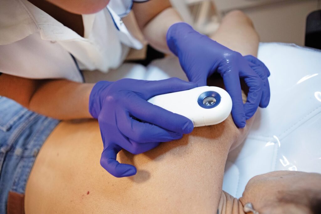
Q: What’s the purpose of that little thingie with the eyepiece that my dermatologist puts on different spots during my skin exam?
Maria Wei, MD, PhD: Great question! That device is a specialized handheld magnifier called a dermatoscope. (“Dermoscopy” refers to the procedure of using a dermatoscope.) This wonderful tool magnifies the skin 10 times larger than what you can see with the naked eye. A dermatoscope uses polarization, which decreases scattered and reflected light. This, essentially, acts as a filter, allowing your doctor to see past the surface of your skin and into the two layers called the epidermis and dermis. Before your physician uses the dermatoscope, they’ll generally first give your skin a thorough head-to-toe visual inspection.
If they see any spots that appear unusual or concerning, they’ll have you remove your makeup or tinted sunscreen and proceed with the dermoscopy exam. Sometimes, your dermatologist will hover the device just over the surface of your skin (often to look at vascular skin changes), but usually they’ll place the scope directly on your skin (or nails) to examine skin changes like warts, moles, fungus and, of course, potentially dangerous conditions like melanoma, squamous cell carcinoma (SCC) and basal cell carcinoma (BCC).
But just because your doctor uses the scope, that does not mean there’s something to worry about. In fact, the vast majority of lesions we look at with the dermatoscope are benign. Because the dermatoscope is so good at helping to definitively identify benign growths, we can avoid many unnecessary biopsies — and the accompanying risk of scarring, bleeding, pain and infection. Plus, fewer needless biopsies means we can also eliminate the uncertainty and anxiety that goes hand-in-hand with waiting to hear if you’ve got skin cancer.
The dermatoscope’s greatest value, though, is in skin cancer detection. I was part of a study published in JAMA Dermatology in 2024 that showed experienced dermatologists (those with over two years of practice) were nearly six times better at identifying melanoma when using dermoscopy compared with visual examination alone. Even more striking, when using dermoscopic imaging, dermatologists were more than 13 times more likely to accurately diagnose melanoma than primary care physicians.
The benefits extend beyond melanoma detection, too: Dermatologists were 2.5 times better at diagnosing nonmelanoma skin cancers — specifically BCCs and SCCs — when using dermoscopy versus standard inspection methods. All of this improved accuracy in skin cancer screening likely leads to earlier detection and better outcomes overall.
While a visual exam without a dermatoscope remains the current standard of care, dermoscopy training is now part of dermatology residency programs nationwide, meaning more physicians will be using this technology. If you’re someone with several melanoma risk factors, like red hair, a family history of the disease, a high mole count or blistering sunburns in your past, you would likely benefit from seeing a specialist who can assess your risk level and determine if you need more frequent monitoring with a dermatoscope. Having your skin examined with a dermatoscope may provide both you and your doctor with greater confidence in your skin health. — Interview by Holly Pevzner
 ABOUT THE EXPERT:
Maria Wei, MD, PhD, is a professor of dermatology at the University of California, San Francisco (UCSF) Helen Diller Family Comprehensive Cancer Center. She’s also a staff physician at the San Francisco VA Health Care System. At UCSF, she leads a laboratory studying skin cancer prevention, melanoma outcomes and the effects of air pollution on the skin.