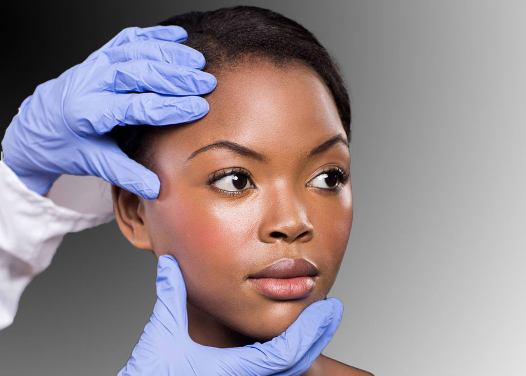
Keep an Eye on Your Skin: If you see anything new, changing or unusual on your skin, trust your instinct and see a dermatologist. Photo: MICHAELJUNG/ISTOCK/GETTY IMAGES PLUS
Q: The new, changing or unusual spots on my skin don’t look like pictures I see online or in brochures. Does that mean I don’t need to worry?
This is a question I get all the time, and the short answer is no — you should not take false comfort from the fact that your lesion doesn’t resemble the ones in photographs. People will often say, “I had a bump that looked nothing like what I saw in the brochure, and it turned out to be skin cancer.” There are many reasons for that. The first is that there are a lot of variations in the way skin cancers look, and they don’t always fit the textbook definitions of a melanoma or a squamous cell carcinoma (SCC) or a basal cell carcinoma (BCC). For example, certain variants of melanoma, including a type called amelanotic melanoma, do not have any pigment; they don’t look like brown spots. Even dermatologists can be fooled.
There are also many unusual variants of BCCs and SCCs that don’t match the examples in textbooks. Then there is the imperfect nature of photography itself: It’s a two-dimensional medium, and a close-up of a classic lesion will not fully mirror a person’s three-dimensional reality. Nor do the photos we currently have reflect the enormous variations that exist in skin tone. A lesion can look dramatically different on someone who’s really dark versus someone who’s extremely pale.
Building a bigger and more diverse archive of photos would certainly help, particularly when it comes to skin cancer in people of color. This is an area that has been neglected medically. There’s a wide misconception — among both people of color and everyone else — that people of color don’t get skin cancer. (They do, albeit at lower rates than the general population.) Therefore, doctors and patients alike don’t look for it, and it can end up being quite advanced by the time it’s diagnosed. The result is a higher mortality rate for melanoma and SCCs in people of color than in the general population.
Lesions in people of color can also be very subtle and easily missed if you’re not really looking for them, often because they’re in areas that aren’t examined carefully, if at all, such as the soles of feet, toenails, mucous membranes and the genital area. And more likely than not, photographs of carcinomas in these places do not appear in skin cancer brochures aimed at a general audience. Most brochures focus on Caucasians. We need to accumulate many more photos of skin cancer in people of color and then create specific brochures that get distributed in their communities. That would do a lot on its own to increase awareness.
In the meantime, my advice for anyone, regardless of complexion, is that if you see something irregular on or under your skin — by which I mean a lesion that is changing in size or color, or is bleeding, flaking or doing anything different — have it examined by a dermatologist. That advice applies even when your lesion looks absolutely nothing like the ones you see in photographs.
View our skin cancer pictures.
— Interview by Lorraine Glennon

About the Expert
Hugh M. Gloster Jr., MD, is a member of Associates in Dermatology, a private practice in Fort Myers, Florida. The coauthor of two medical textbooks, he specializes in cutaneous oncology, Mohs surgery and reconstruction.


